
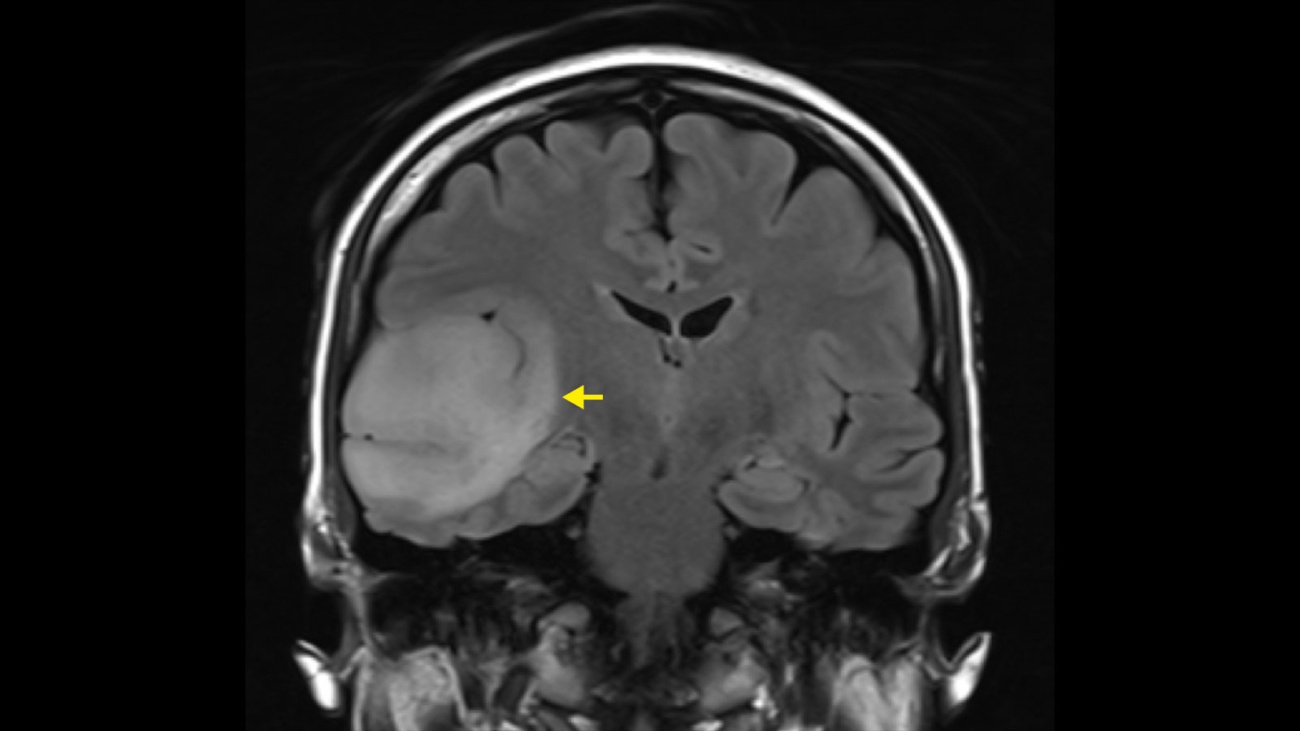
Surgical planning for brain disorders depends on clear, detailed imaging that helps the care team understand what is happening and where it is located. Interventional radiology supports this process by providing advanced, image-guided techniques that clarify anatomy and assess blood vessels. Here’s a closer look at the role of neuroradiology in surgical planning for brain disorders:
Mapping Targets
One of the fundamental tasks in surgical planning is defining where the problem lies and identifying healthy tissue that must remain intact. Surgeons need to know the precise boundaries of a tumor or lesion so they can remove it completely while protecting surrounding areas. Interventional radiology techniques, such as functional MRI (fMRI), enable specialists to map key brain regions that control speech, movement, and vision. These key areas are often referred to as “no-go zones”, and avoiding them prevents long-term damage to a patient’s quality of life. Detailed mapping helps the surgical team determine the best angle of approach.
Understanding Blood Flow
The brain is filled with a complex network of blood vessels, and disrupting them can cause serious complications. Neuroradiologists use specific imaging modalities to visualize this network and identify potential risks to arteries or veins. Here are several ways doctors assess blood flow to prepare for surgery:
- Angiography: This technique uses contrast dye and X-rays to create a clear picture of the blood vessels, showing exactly how blood travels through the brain.
- Perfusion Imaging: This method quantifies the blood flow to different regions of brain tissue, identifying areas that may be at risk during the procedure.
- Vessel Mapping: Creating a digital map of major arteries and veins enables the surgeon to plan an incision path that avoids injury to major blood supplies.
- Flow Dynamics Analysis: Doctors analyze the speed and pressure of blood flow, which is helpful when dealing with aneurysms or other vascular disorders.
Understanding these vascular patterns is helpful for minimizing blood loss and preventing strokes during the operation.
Using Image-Guided Procedures
Modern technology allows for interventional radiology techniques that reduce physical impact on the patient. Instead of large incisions, doctors can often use small openings and guide instruments using real-time images. Here are some common ways imaging supports these procedures:
- Stereotactic Navigation: This tracks surgical instruments in real-time against the patient’s pre-operative scans.
- Intraoperative MRI: Some operating rooms are equipped with MRI machines, enabling surgeons to take new images during surgery to assess surgical progress.
- Endovascular Treatments: Surgeons guide thin tubes, called catheters, through blood vessels to treat conditions from the inside, often avoiding open brain surgery entirely.
- Biopsy Guidance: Imaging helps doctors guide a needle to a precise location to obtain a tissue sample for diagnosis.
These interventional radiology procedures rely heavily on the expertise of the neuroradiology team to maintain accuracy.
Providing Real-Time Guidance
The role of interventional radiology does not end once the planning phase is complete. During surgery, real-time imaging provides continuous feedback to the surgeon, serving as a continuous verification tool. If the brain shifts slightly during the procedure, the imaging system updates to reflect the new position.
This immediate feedback loop enables the surgeon to adjust their movements instantly, maintaining high precision even as conditions change. After surgery, post-operative imaging is fundamental for assessing immediate outcomes. It also confirms that the target, such as a tumor or obstruction, was successfully treated in accordance with the plan.
Schedule Your Interventional Radiology Consultation
Neuroradiology plays a key role in making brain surgery safe and precise through detailed planning and guidance. These tools provide surgeons with the information they need to navigate the delicate anatomy of the brain. The use of real-time imaging makes sure that accuracy is maintained from the first incision to the final check. If you or a loved one requires specialized care, contact a trusted interventional radiology specialist near you to schedule your consultation today.